Exosome: The Rising Star in Drug Delivery and Dermatologic Therapy Listen with ReadSpeaker Our expertise

Exosome: The Rising Star in Drug Delivery and Dermatologic Therapy

Exosomes are microvesicles released by cells in both physiological and pathological situations. They are surrounded by a lipid bilayer with proteins derived from the origin cell and contain a variety of molecules including nucleic acids.

They represent an emerging mechanism of intercellular communication, and they play an important role in the pathogenesis of cancer, stimulating the proliferation and aggressiveness of cancer cells, inducing a microenvironment favorable to tumor development, and controlling immune responses.

Because of the growing understanding of the potential implications of extracellular vesicles (EV) in the development of malignancies, research on exosomes, and their role as a diagnostic and therapeutic tool, constitutes nowadays a very exciting and promising field.

Exosome role Details and references
Cell–cell communication Exosomes can participate in autocrine, paracrine, or endocrine communication reaching their target cells via systemic or local circulation. They are important participants in cell communication including cell migration, proliferation, and senescence.
Immune response The cells of the immune system are known to release exosomes. Exosomes mediate immune modulation, both immunosuppression and immunostimulation.
Signal transduction Exosomes enable intercellular communication between various types of cells, regulating gene expressions and cellular signaling pathways of recipient cells by delivering their components, such as specific lipids, proteins, and RNAs. Certain lipid components including sphingomyelin, cholesterol, and ceramides have been involved in signaling; phosphatidylinositol-3-phosphate (PI3P) is also known to participate in regulating cell signaling. The presence of multiple kinds of signaling molecules: lipids, proteins, and RNAs results in rapid signal changes in the target cell.
Material (cargo) transport Exosomes transport their constituents’ proteins, nucleic acids, lipids, and metabolites between cells, both in the close vicinity of the parent cell and at distant sites in the body carried by biofluids. It has been reported that RNA cargo of exosomes can modify gene expression in recipient cells.
Pathogenesis Viruses are known to make use of exosome biogenesis pathways to release a variety of pathogenic factors. Thus, several pathogen-derived components have been detected on exosomes after infection. These include, e.g., human immunodeficiency virus, Epstein–Barr virus, cytomegalovirus, hepatitis C virus, and herpes simplex virus. Exosomes play multiple roles in the progression of cancer via various communication pathways. Exosomes are more often released by tumor cells than by healthy ones and facilitate communication within the tumor microenvironment.
Blood–brain communication Exosomes can cross the BBB in both directions: from the brain to the bloodstream and from the blood to the CNS. Moreover, exosomes can interact with the BBB, leading to changes in the barrier’s properties.
Target cell delivery The delivery of cargos such as bioactive RNAs, proteins, metabolites, and/or lipid makes the capture of exosomes by target cells of vital importance in a variety of key biological processes such as angiogenesis, bone development, and cell migration.


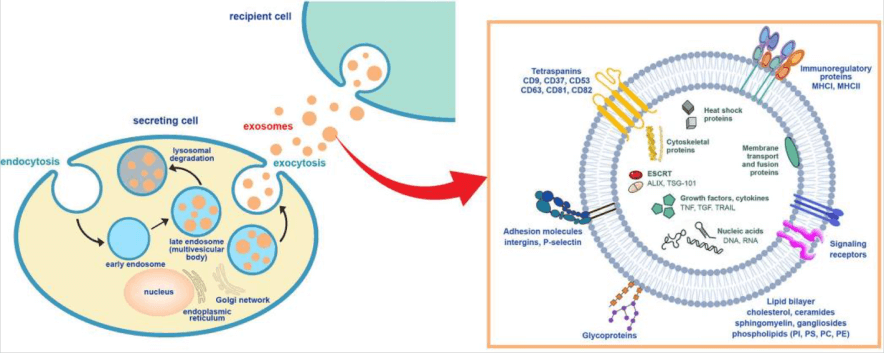
Scheme of exosome biogenesis and secretion. The inset exemplifies the molecular constituents of the exosomes.


Source

Exosomes have recently been gaining attention in the cosmetic world. Used in topical creams, serums, and masks, exosomes have been found to have several therapeutic and anti-aging benefits. Exosomes have been found to be beneficial for skin care, as they are filled with proteins, lipids, and other molecules that can help to promote healing, hydration, and the protection of the skin.


These molecules can help to boost collagen production, reduce inflammation, and protect the skin from environmental stressors. Additionally, exosomes can help to increase the efficacy of other active ingredients, such as hyaluronic acid, peptides, and antioxidants.


Figure 1 depicts Adipose stem cell derived-condition media (ASC-CM), Bone marrow stem cell derived (BMSC)-exosomes, decreased reactive oxygen species (ROS), as well as TNF-α and increased TGF-β, resulting in higher MMP-1 and pro-collagen type I. This led to the increase of collagen synthesis, the improvement of elasticity, and the reduction of wrinkles, an effective anti-aging therapy. Filled with molecules that help stimulate collagen production, exosomes can help to reduce wrinkles and fine lines.


Additionally, exosomes can help to repair skin damage such as sun damage and acne scars. Exosomal proteins and lipids can help to plump and hydrate the skin, which can help to improve skin texture. Ingredients of exosomes such as cytokines, nucleic acids, proteins, and other bioactive compounds can also help to protect the skin from environmental stressors and reduce the appearance of dark spots and other discoloration. With their ability to help improve skin tone, texture, and appearance, exosomes provide several promising therapeutic and anti-aging benefits.

Cell therapy practices date back to the 19th century and continue to expand on investigational and investment grounds. Cell therapy includes stem cell- and non–stem cell-based, unicellular, and multicellular therapies, with different immunophenotypic profiles, isolation techniques, mechanisms of action, and regulatory levels.


Following the steps of their predecessor cell therapies that have become established or commercialized, investigational, and premarket approval-exempt cell therapies continue to provide patients with promising therapeutic benefits in different disease areas.


Stem cells mainly include embryonic stem cells (ESC), pluripotent stem cells (iPS), Epiblast-derived stem cells (Epi-SC), and adult tissue stem cells. ESC are pluripotent, being able to differentiate into the ectoderm, mesoderm, and endoderm (Evans and Kaufman, 1981; Martin, 1981). However, there are ethical issues when using human embryos, and ESC transplants into mouse models can result in malignant tumors. Further challenges facing research in the use of ESC are how to reduce contamination and prevent cancer risk.


In contrast to stem cells, exosomes cannot self-replicate, eliminating concerns about potential tumor formation after stem cell transplantation. Exosomes are also stable enough for long-term frozen storage and storage at room temperature after lyophilization. Their small size further allows sterilization by filtration. In addition, exosomes can be administered by several routes; for example, nebulized or lyophilized lung stem cell-derived exosomes can be administrated by inhalation to treat lung diseases.


Moreover, their hydrophilic lumen and phospholipid bilayer containing membrane proteins can be engineered and modified to display molecules or for drug loading; lung stem cell-derived exosomes were recently decorated with the receptor-binding domain of recombinant SARS-CoV-2 as an inhalable COVID-19 vaccine. In 2020, the first clinical trial (NCT04592484) of an engineered exosome therapy (exoSTING8) was launched by Codiak Biosciences for the treatment of multiple solid tumors, indicating that engineering exosomes may be a future direction for therapeutic applications of stem cell-derived exosomes.

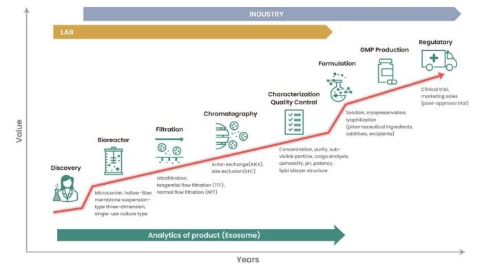
Upstream cell culture. To date, exosomes have been produced from stem cells or immune cells in clinical trials, including bone marrow mesenchymal stem cells, adipose tissue-derived stem cells, and monocyte-derived dendritic cells (Mendt et al., 2019; Näslund et al., 2013; Zhang et al., 2020a).


A flask-based static system in the cell culture chamber can be used for lab-scale culture of these originator cells; however, bioreactor systems, such as microcarriers, hollow-fiber membranes, and microfiber-bed types, are commonly used for large-scale culture (Chen et al., 2011; Valkama et al., 2018). Attempts to increase culture capacity are ongoing; however, increasing the productivity of exosomes generated from adherent cells, such as stem cells, is very limited.


Downstream exosome purification. Exosome size ranges from 50 to 200 nm, and many exosome purification methods separate exosomes from various EVs based on their size and density. One of the most used methods, normal flow filtration (NFF), a molecular weight cutoff system based on membrane pore size, utilizes vacuum or centrifugal forces to filter the culture media.


NFF is often used in lab-scale early development to purify exosomes from cell culture media. However, its application to industrial-scale bioprocesses is highly limited due to prolonged processing time and limited scalability. In addition, these purification methods are inadequate for producing high-purity exosomes. Therefore, alternative purification steps that yield high purity are required for further drug development.


Source

Many considerations should be made to optimize exosome quality, such as optimizing the originator cell line, culture and harvest conditions, separation and purification methods, and stability (Théry et al., 2006; Zhang et al., 2020b). In this section, we focus our discussion on the analytical methods and criteria for exosome quality control.


Exosome characteristics must be analyzed to determine the quality of the produced exosomes. General characterization includes measuring exosome particle number per volume using nanoparticle tracking analysis (NTA), the presence of positive/negative exosome markers using immunoblotting, and the size and structure of the lipid bilayer using cryo-electron microscopy (Cryo-EM) (Table 1) (Gurunathan et al., 2019; Kurian et al., 2021; Szatanek et al., 2017; Thery et al., 2018).


Assays detecting originator cell-specific markers or common exosome markers, such as proteins, lipids, or nucleic acids, can be used to analyze exosome identity. In most cases, exosome-specific proteins have been identified using western blotting or FACS with a specific antibody (Yang et al., 2019). Recently, exosomes have been highlighted as potential drug carriers (Song et al., 2021; Yim et al., 2016). These therapeutic cargo molecules require quantitative or qualitative characterization.


In addition, other components that comprise exosomes should be profiled, because they contain many different proteins, lipids, and nucleic acids from their host cells (Choi et al., 2013; 2020). Exosome profiling results can be used to predict genotoxicity and/or carcinogenicity by analyzing the proteins or nucleic acids that are responsible for inducing such effects.


The safety profile of a drug is one of the most critical aspects of drug discovery. Thorough profiling of exosomes, including proteins, lipids, and nucleic acids, as well as conducting animal toxicology studies, will be critical for predicting any unforeseen safety issues.

Reliable extracellular vesicles isolation and characterization is fundamental to drug discovery research and essential in disease detection and treatment. Rapid identification of elevated exosome concentrations can indicate the onset or progression of the disease, while harnessing EVs unique properties as drug delivery vehicles provide a promising alternative route to combat disease.


However, when extracted from bodily fluids in response to specific conditions, EVs are highly heterogeneous, making the characterization of these invisible bubbles uniquely challenging.

NanoSight instruments, the first to incorporate Nanoparticle Tracking Analysis (NTA) have been assisting exosome researchers over the decade offering high-resolution size and concentration characterization of these complex samples. The newest instrument, NanoSight Pro packed with intelligence and enhanced sensitivity is an ideal toolbox for every lab. New software, powered by machine learning, takes NTA to the next level providing easy, quick, reliable data.

In addition, sensitive detection provides very detailed information about samples in both the light scatter and fluorescence modes. By enabling specific detection of biomarkers and cargo, you get a step closer to understanding the personality of your extracellular vesicles to decode their message in the bottle.

Sources:

Ruethaitip Tiratrakulvichaya

About the author

Ruethaitip Tiratrakulvichaya has been with DKSH in Thailand since 2009. As the Application Manager for the Malvern product range, she is responsible for technical and application support across Southeast Asia.

With a background in food science and agroindustry, she is experienced in delivering training to both internal colleagues and external customers on how to operate and obtain the best data.

Ruethaitip has extensive working knowledge in material characterization techniques including laser diffraction, dynamic light scattering, micro-calorimetry, size exclusion chromatography, and morphological property.